
Ultrasound Tissue Characterization
Abstract
Among the non-invasive imaging modalities, ultrasound imaging is, probably, the most widespread technique. The main reason of its success is that it provides a low-cost way to help diagnosing and can be used in many medical applications. However, ultrasonic (US) images are characterized by the presence of a peculiar granular pattern, the so-called speckle.
This term was adopted from the field of laser optics in the early sixties due to the similarity of the patterns between laser optics and ultrasonics. Although the nature of the speckle in US images stems from a different phenomena, there still share some similarities. Both patterns come from the random interference of many coherent wave components reflected from different microscopic elements. In the case of US, the volume, the number of effective scatterers, and the acquisition process contribute to the formation of speckle.
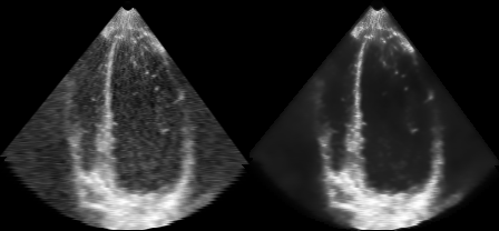
The analysis of backscattered echo from tissues needs a proper description of the US signals. For this purpose, and due to the random nature of the speckle, several statistical models have been proposed in the literature. This characterization can be used either for segmentation, classification or for filtering the speckle itself. The latter usually considers the speckle as an undesired consequence, since it degrades resolution and adds spatial noise to the image. Thus, filtering is commonly applied as a preprocessing step for further segmentation of regions of interest or to extract relevant measures for physiological analysis.
The statistical description of US signals provide an important information of the backscattered echo from tissues. The parameters of the statistical models allow identifying the features of these tissues and provides important descriptors for classification. Some of the filtering algorithms relay on a Bayesian approach where an accurate statistical model becomes necessary. As a consequence, modeling the amplitude statistics of US signals has been a very active area. Thus, several statistical models have been proposed in last decades.
Probably the most well-known is the Rayleigh model, which is a one-parameter distribution which describes the so-called fully formed (or developed) speckle. This probabilistic distribution of the amplitude of US signals describes the behavior of speckle when a high number of effective scatterers is present in the resolution cell. However, real images show a deviation from this model. This non-Rayleigh behavior can be due to a small number of scatterers in the resolution cell or when there are some dominant components in the cell. The most commonly accepted distributions that try to model non-Rayleigh distributions are the Rice (fully resolved speckle), K (partially formed speckle) and Homodyned K (partially resolved speckle).
Although those models are based on physical assumptions of the backscattering process, some other distributions have proven to provide a good performance on real images. This is the case of Gamma and Nakagami distributions. The first is proposed as a two-parameter distribution that describes the result of interpolated/filtered fully formed speckle and also has shown good results in empirical tests among other distributions. The Nakagami distribution proposed by Shankar for the case of US characterization is also a two-parameter distribution which generalizes the Rayleigh distribution. This distribution was adopted from the models proposed to describe the statistics of the returned radar echo.
The capability of the Nakagami distribution to model the backscattering from tissues for fully resolved and fully formed speckle render it one of the most commonly accepted model for tissue characterization. However, the tails of the probabilistic density functions of Nakagami, K, Rayleigh or Gamma do not show the impulsive response of speckle which originates heavier tails. In order to describe this impulsive response, a ge\-neralized Nakagami distribution was proposed by Shankar. This is a three-parameter model which has shown a better behavior than the Nakagami or Rayleigh, an expected result since it is a generalization of the other models. However, the generalized Nakagami distribution does not have closed-form Maximum Likelihood (ML) estimates and, thus, it makes their use difficult.
In summary, there is no agreement on the probabilistic distributions that model the different nature of tissues in US. This is due to the complex composition of tissues that can show combinations of different kinds of speckle in the resolution cell. Besides, the probabilistic nature of speckle is affected by every acquisition step before the image is fully acquired.
In this research we intend to characterize the random nature of tissues in US imaging through the different steps of the acquisition process. To that end, it will be necessary to develop some mathematical tools to characterize probabilistic distributions of models with an increasing complexity along the acquisition process of the image. Once the tissues and noise are properly characterized, we propose some theoretical and practical contributions to the field of US and MRI imaging.
Publications:
Journal Publications
- G. Vegas-Sánchez-Ferrero, S. Aja-Fernández, M. Martín-Fernández, C. Palencia, “A direct calculation of moments of the Sample Variance”, Mathematics and Computers in Simulation, Volume 82, Issue 5, January, Pages 790-804. 2012.
- Gonzalo Vegas Sánchez-Ferrero, José Seabra, Santiago Aja-Fernández, Marcos Martín-Fernández, César Palencia, Joao Sanches. "Gamma Mixture Classifier for Plaque Detection in Intravascular Ultrasonic Images". IEEE Transactions on Ultrasonics, Ferroelectrics, and Frequency Control. vol.61, no.1, pp.44,61, January 2014.
- Gonzalo Vegas-Sánchez-Ferrero, Santiago Aja-Fernández, César Palencia, Marcos Martín-Fernández. "Generalized Gamma Mixture Model for Ultrasonic Tissue Characterization". Computational and Mathematical Methods in Medicine. Article ID 481923, 25. 2013.
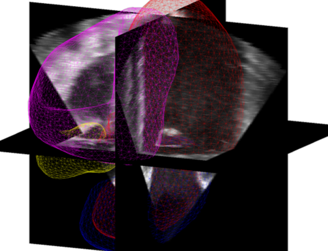
- A. Haak, G. Vegas-Sánchez-Ferrero, H. W. Mulder, B. Ren, H. A. Kirisli, C. Metz, G. van Burken, M. van Stralen, J. P. W: Pluim, A. F.W. van der Steen, T. van Walsum, J. G. Bosch, "Segmentation of multiple heart cavities in 3D transesophageal ultrasound images", IEEE Transactions on Ultrasonics, Ferroelectrics, and Frequency Control. Accepted.
- G. Ramos-Llordén, G. Vegas-Sánchez-Ferrero, M. Martín-Fernández, S. Aja-Fernández, C. Alberola-López, "Anisotropic diffusion filter with memory based on speckle statistics for medical images". IEEE Transactions on Image Processing. Accepted.
Conference Publications
- Gonzalo Vegas-Sánchez-Ferrero, Diego Martín-Martínez, Santiago Aja-Fernández, César Palencia “On the Influence of Interpolation on Probabilistic Models for Ultrasonic Images”, Symposium on Biomedical Imaging: From nano to macro (ISBI). Rotterdam (Netherlands). 2010.
- Santiago Aja-Fernandez, Gonzalo Vegas-Sanchez-Ferrero, Miguel Angel Martin-Fernandez, “Soft Thresholding for Medical Image Segmentation”, EMBC, Argentina Jul 2010.
- Gonzalo Vegas-Sánchez-Ferrero, Santiago Aja Fernández, Marcos Martín Fernández, Alejandro Frangi, César Palencia. “Probabilistic-Driven Oriented Speckle Reducing Anisotropic Diffusion with Application to Cardiac Ultrasonic Images”. International Conference on Medical Image Computing and Computer Assisted Intervention (MICCAI), Beiging, Sep 2010.
- Gonzalo Vegas-Sánchez-Ferrero, Diego Martin-Martinez, Pablo Casaseca-de-la-Higuera, Lucilio Cordero-Grande, Santiago Aja Fernández, Marcos Martín Fernández, César Palencia. “Realistic Log-Compressed Law for Ultrasound Image Recovery”. 18th IEEE International Conference on Image Processing. 2011.
- A.H. Curiale, G. Vegas Sánchez-Ferrero, T. Pérez-Sanz, S. Aja-Fernández, “Cuantificación de la insuficiencia mitral funcional mediante el tensor de esfuerzo y la velocidad del miocardio”. Congreso Anual de la Sociedad Española de Ingeniería Biomédica (CASEIB), Cáceres, Spain. 2011.
- G. Vegas Sánchez-Ferrero, A. Tristán Vega, S. Aja-Fernández, M. Martín-Fernández, C. Palencia, R. Deriche. “Anisotropic LMMSE denoising of MRI based on statistical tissue models”. Symposium on Biomedical Imaging: From nano to macro (ISBI). Barcelona (Spain). 2012.
- G. Vegas Sánchez-Ferrero, F. Simmross Watemberg, M. Martín Fernández, C. Palencia, C. Alberola López, "Caracterización de Speckle con Modelos de Cola Pesada", Congreso Anual de la Sociedad Española de Ingeniería Biomédica (CASEIB). San Sebastián 2012.
- G. Ramos Llordén, G. Vegas Sánchez-Ferrero, S. Aja Fernández, M. Martín Fernández, C. Alberola López, "Filtro de Difusión Anisótropo con Memoria basado en Modelos Probabilísticos para Imágenes Intravasculares y Cardíacas", Congreso Anual de la Sociedad Española de Ingeniería Biomédica (CASEIB). San Sebastián 2012
- Zeynettin Akkus, Johan G. Bosch, G. Vegas Sánchez-Ferrero, Diego D. B. Carvalho, Guillaume Renaud, Stijn C. H. van den Oord, Gerrit L. ten Kate, Arend F. L. Schinkel, Nico de Jong, Antonius F. W. van der Steen, "Statistical segmentation of carotid plaque neovascularization", Proc. SPIE 8675, Medical Imaging 2013: Ultrasonic Imaging, Tomography, and Therapy, 867506. 2013.
- Ariel Hernán Curiale, Gonzalo Vegas-Sanchez-Ferrero, Teresa Miriam Pérez-Sanz, Santiago Aja-Fernandez "Strain rate tensor estimation from Echocardiography for quantitative assessment of Functional Mitral Regurgitation", on Biomedical Imaging: From nano to macro (ISBI). San Francisco, CA, USA. Apr. 2013.
- Ariel Hernan-Curiale, Gonzalo Vegas-Sanchez-Ferrero, and Santiago Aja-Fernandez, "Speckle Tracking in Interpolated Echocardiography to Estimate Heart Motion". International Conference on Functional Imaging and Modeling of the Heart, Jun. 2013.
- Alexander Haak, Gonzalo Vegas-Sánchez-Ferrero, Harriët H. Mulder, Hortense A. Kirisli, Nora Baka, Coert Metz, Stefan Klein, Ben Ren, Gerard van Burken, Anton F. W. van der Steen, Josien P. W. Pluim, Theo van Walsum, Johan G. Bosch: "Segmentation of 3D Transesophageal Echocardiograms by Multi-cavity Active Shape Model and Gamma Mixture Model". AE-CAI, volume 8090 of LNCS, page 19-26. Springer, 2013.
- G Ramos-Llordén, G. Vegas-Sánchez-Ferrero, S Aja-Fernández, M Martín-Fernández, C Alberola-López. "Fast Anisotropic Speckle Filter for Ultrasound Medical Images". XIII Mediterranean Conference on Medical and Biological Engineering and Computing 2013. IFMBE Proceedings Volume 41, pp 253-256. 2014.
- Haak, A. ; Vegas-Sanchez-Ferrero, G. ; Mulder, H.H. ; Kirisli, H.A. ; Baka, N. ; Metz, C. ; Klein, S. ; Ben Ren ; van Burken, G. ; Pluim, J.P.W. ; van der Steen, A.F.W. ; van Walsum, T. ; Bosch, J.G., “Simultaneous segmentation of multiple heart cavities in 3D transesophageal echocardiograms“,Ultrasonics Symposium (IUS), 2013 IEEE International, 2013.
- Haak, A. ; Mulder, H.W. ; Ren, B. ; Vegas-Sanchez-Ferrero, G. ; van Burken, G. ; van der Steen, A.F.W. ; van Stralen, M. ; Pluim, J.P.W. ; van Walsum, T. ; Bosch, J.G. “Segmentation of multiple heart cavities in wide-view fused 3D transesophageal echocardiograms”, Ultrasonics Symposium (IUS), 2014 IEEE International, 2014.
Book Chapter
- G. Vegas-Sánchez-Ferrero, M. Martín-Fernández, J. Miguel Sanches "A Gamma Mixture Model for IVUS Imaging", Multi-Modality Atherosclerosis Imaging and Diagnosis. Editors: Luca Saba, João Miguel Sanches, Luís Mendes Pedro, Jasjit S. Suri ISBN: 978-1-4614-7424-1 (Print) 978-1-4614-7425-8 (Online), Pages 155-171. 2014.
Patents
- Gonzalo Vegas Sánchez-Ferrero, Marcos Martín Fernández, Santiago Aja Fernández, Carlos Alberola López. “Método de Filtrado y Realce de Imágenes Ultrasónicas 2D, 3D y 3D+t.” OEPM Madrid, Num: P201430673. Under formal evaluation, 2014
ITK Code
- A. H. Curiale, G. Vegas-Sánchez-Ferrero and S. Aja-Fernández, “Probabilistic Tissue Characterization for Ultrasound Images,” Insight Journal (2015), http://hdl.handle.net/10380/3517. http://www.insight-journal.org/browse/publication/955